Nobel Prize in Chemistry 2014 - how can we see the hidden depth with an optical microscope?
A microscope allows you to see very small species. the microscope has its limitations – if you try to look at an object that is less than 1/5000 of a millimeter, you cannot achieve a contrasting picture. See how scientist developed new tool to overcome this limitation and won the Nobel Prize
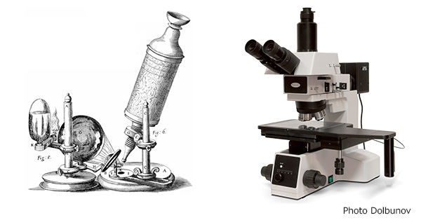
Classical microscopy
Humans’ wish to harness six senses has existed since civilization started. People always wanted to know more, see more, and hear better. The vision developed as the sense giving more information than other senses. Especially in science, with its very complicated subjects, visual representation of different objects is very important. We want to know the truth about every object we see. That’s why it is very important to get information which is not seen by normal vision. So the microscope was developed. But the microscope has its limitations – if you try to look at an object that is less than 1/5000 of a millimeter, you cannot achieve a contrasting picture.
The failure to get an image with a very high resolution with an optical microscope is called the diffraction limit. What is the reason for this limit to occur? Our eye as well as a microscope can see something only if it catches light reflected from an object. The light is a wave. If the object is much less in size than the size of a wave, directed to an object, than the wave we can then walk around this object.
Limitations of classical microscopy
As an example, you may remember your last boat trip on a sea: waves coming from the sea go around your boat and do not reflect from it. However, the light being a wave with less wavelength, reflects from your boat and you can see it with an untrained eye. The same mechanism works in microscopy: we need to use very small waves to see small objects. Optical light has its limits. When you take an object smaller than the smallest optical light wave, you cannot see this thin object with optical microscopy working on a principle of light reflection.
See a 2-minute video about diffraction:
How can we see a single molecule?
If you find a way to use a wavelength comparable to the object size, you can win the Nobel Prize. The three scientists Eric Betzig, Stefan W. Hell, and William E. Moerner developed the tool that can assist you to see molecules less in size than the “size of a light wave” (wavelength).
Please remember the last time you took a photo for a passport. You were asked to make a photo using a white background due to the contrast between your face and the color of the background. The same way can be used for molecules. We need a very good signal from every single molecule and it should be contrasted with signals from other molecules. But we cannot ask molecules to take their photo on a white background. What we can do, however, is to ask molecules that are close to another, to send us signals in different time intervals, so we can distinguish them from the average background.
Some molecules especially which occur in a living cell (proteins) demonstrate a property called fluorescence. The fluorescence means that chemical substances emit light being under some external action – chemical reaction or a different light source. Using fluorescence induced by a light one may obtain a picture of single protein molecules. For that purpose, you should irradiate only a very thin part of an object. That was the reason for Stefan Hell to use lasers for the irradiation of samples. The contrast was still lacking. If you irradiate some point at a sample with a laser, you cannot target the single molecule. Different molecules close to one another start to fluorescence and we don’t get a contrasted picture of a single protein.
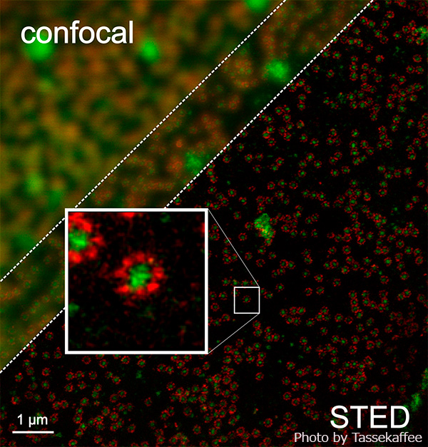
Super-microscopy: STED
To overcome this obstacle, the second tool was developed: STED (stimulation emission depletions). That tool allows you to see fluorescence in a very thin spot using two laser beams: one for enhancing fluorescence in the center of that beam with the size of nanometers. While another beam used for the depression of fluorescence in the nearest regions around the nm-size spot. With this complicated technique we can force one protein molecule to send us a fluorescence signal while other molecules are silent – emitting no radiation.When you get an image from one spot, you may continue to scan the sample with a double laser beam, and get a full image of the object in the scanned area. This information will contain a single protein.
The Nobel Prize was given to three different scientists working on a similar theme. The second scientist, W.E.Moerner, was first to measure the light absorption of a single molecule while Eric Betzig invented a way to distinguish molecules of different colors even if they lay at a distance just a few nanometers apart. This invention is used for imaging how a disease develops in a living cell and can bring a huge impact to the fundamental understanding of the molecular mechanisms inside living organisms.
Perspective application of STED microscopy are not limited to biology. There are some perspective applications in chemistry, especially in catalysis.
This article used materials from the official website of the Nobel Prize.
See also
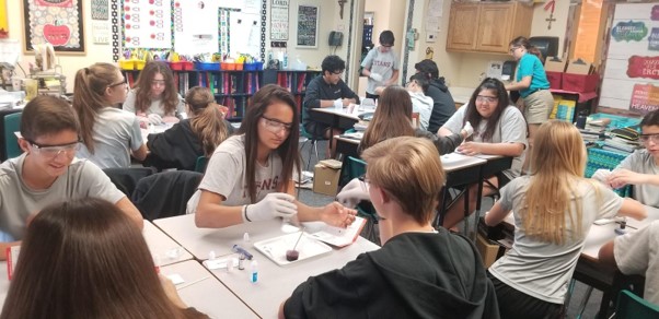
CASE STUDY - 8th Grade students at St Timothy's Catholic School use MEL Chemistry to enhance their science lessons
Saint Timothy Catholic School in Mesa is committed to promoting academic excellence in each child it looks after. They encourage self-discipline, self-respect, and respect for others. They understand the importance of engaging students in a comprehensive and relevant curriculum. As a result, the middle school science teacher from St. Timothy Catholic School is using MEL Chemistry subscriptions to enhance and expand their range of learning activities.
CASE STUDY - MEL Chemistry allowing pupils to reach their full potential
The Empower Learning Center is the Alternative Learning Program (ALP) within the Hinckley-Finlayson School District. They offer non-traditional education options for students ages 16-21 in their daytime program, night school for traditional high school students who need to make up credits, and night school for adults 18 and older who would like to complete their diploma or equivalency.
The school was seeking engaging, hands-on chemistry kits to make their science classes more interactive, and to help their students understand key science concepts and achieve their full potential in chemistry.
CASE STUDY - MEL Chemistry at Lund International School, Sweden
Emma Taylor, a science teacher at Lund International School (Sweden), has chosen MEL Chemistry sets as the best option for her students’ science classes. In Lund International School, all programmes are taught in English, and having chemistry sets in English are a great asset to accompany science classes.
Here, Emma shares her experience of how MEL Chemistry sets improved her students’ comprehension and understanding of science concepts.